In the realm of medical imaging, positron emission tomography (PET) has emerged as a revolutionary technique, offering unparalleled insights into the body’s metabolic processes. At the heart of this technology lies the ingenious design of PET microchips, miniature marvels that enable the detection and visualization of radioactive tracers within the body. Understanding how PET microchip work means embarking on a captivating journey into the intricate workings of PET microchips and uncover the principles that empower them to transform medical diagnosis and treatment.
Unveiling the Essence of PET Microchips
PET microchips, also known as PET detectors, are the fundamental building blocks of PET scanners. These meticulously crafted devices are responsible for capturing the annihilation photons emitted by radioactive tracers, converting them into electrical signals that can be analyzed to produce a detailed image of the body’s metabolic activity.
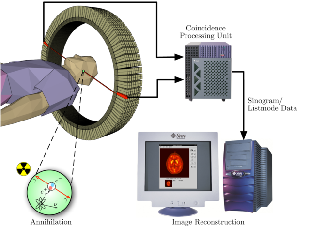
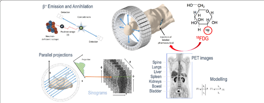
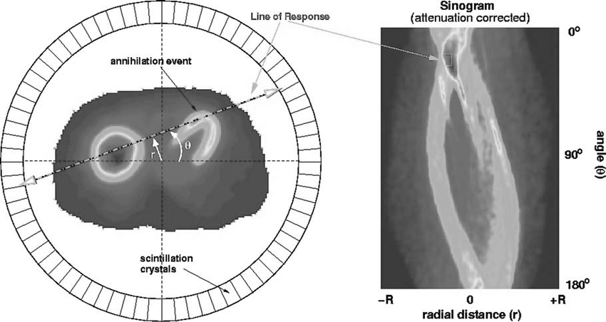
Imagine a PET microchip as a tiny guardian angel, diligently monitoring the body’s inner workings. As radioactive tracers traverse the bloodstream, they encounter areas of high metabolic activity, such as tumors or regions of the brain with increased activity. Upon reaching these locations, the tracers decay, emitting positrons. These positrons, like fleeting sparks, collide with electrons, their paths converging in a dance of annihilation. It is during this annihilation that a burst of energy, in the form of annihilation photons, is released.
Delving into the PET Microchip Architecture
A PET microchip comprises a scintillator crystal, a photomultiplier tube (PMT), and associated electronics. The scintillator crystal, typically composed of materials like bismuth germanate (BGO) or cerium-doped lutetium yttrium orthosilicate (LYSO), serves as the primary interaction site for annihilation photons. Upon striking the crystal, these photons generate a cascade of scintillation light particles.
Picture the scintillator crystal as a miniature light show. As annihilation photons collide with atoms within the crystal, they impart their energy, causing electrons to jump to higher energy states. When these electrons return to their ground states, they release their energy in the form of scintillation light. This cascade of light particles, much like a trail of breadcrumbs, guides the annihilation photons towards the next stage of the detection process. This is a breakdown of how PET microchip work.
Harnessing the Power of Scintillation Light
how PET microchip work won’t be complete without the scintillation light produced within the crystal is then guided towards the PMT, a highly sensitive light detector. Inside the PMT, the photons are converted into electrons through a process known as photoemission. These electrons are subsequently amplified through a series of dynodes, resulting in a measurable electrical signal.
Imagine the PMT as a light-amplifying powerhouse. As scintillation photons enter the PMT, they strike a photocathode, releasing electrons. These electrons are then accelerated towards a series of dynodes, each coated with a material that emits more electrons when struck by an incoming electron. This cascading effect, known as electron multiplication, amplifies the initial signal, producing a measurable electrical pulse.
Transforming Signals into Images
Understanding how PET microchip work means that the electrical signals generated by the PMT are then processed by sophisticated electronics, which extract the timing and amplitude information associated with each photon detection event. This data, meticulously compiled, forms the basis for reconstructing a detailed image of the body’s metabolic activity.
Envision the electronics as a meticulous data analyst, meticulously organizing and interpreting the electrical signals. The timing information reveals the location of each annihilation event, while the amplitude information provides insights into the energy of the photons, which in turn relates to the metabolic activity in that region. This intricate dance of data analysis transforms a barrage of signals into a comprehensive image of the body’s metabolic landscape.
The Role of Radioactive Tracers in PET Imaging
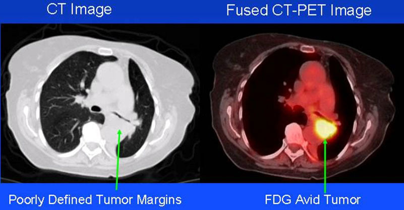
Radioactive tracers, such as 18F-fluorodeoxyglucose (FDG), play a pivotal role in PET imaging. These substances are injected into the bloodstream and accumulate in areas of high metabolic activity, such as tumors or regions of the brain with increased activity. When FDG decays, it emits positrons, which annihilate with electrons, producing the annihilation photons detected by PET microchips.
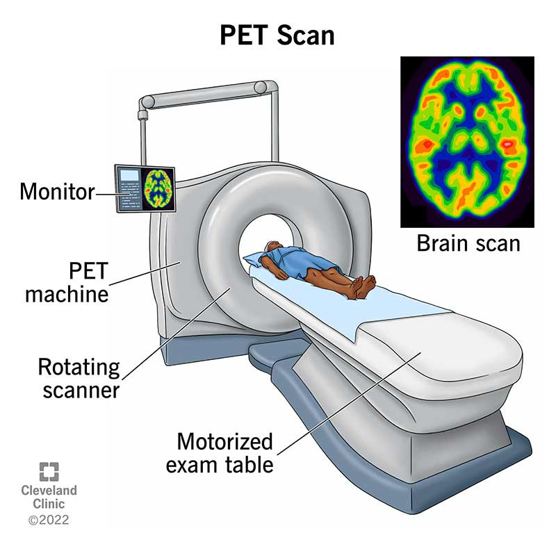
Consider FDG as a metabolic beacon, guiding PET microchips towards areas of interest. As FDG molecules circulate through the bloodstream, they are selectively taken up by cells with high glucose demand, such as tumors or regions of active brain function. Upon reaching these locations, FDG decays, emitting positrons that serve as landmarks for PET microchips, revealing the body’s metabolic hotspots. See image above.
The Impact of PET Microchips on Medical Imaging
PET microchips have revolutionized medical imaging, providing invaluable insights into a wide range of conditions, including cancer, heart disease, and neurological disorders. By enabling the visualization of metabolic activity, PET has transformed the diagnosis and monitoring of these diseases, leading to improved patient outcomes.
PET microchips have emerged as game-changers in medical imaging, offering a unique window into the body’s metabolic processes.
Conclusion: A Glimpse into the Future of PET Microchip Technology
The future of PET microchip technology holds immense promise, with advancements in scintillator materials, PMTs, and electronics paving the way for even more sensitive, faster, and affordable PET scanners. These innovations will undoubtedly expand the reach of PET imaging, making it an even more indispensable tool for diagnosing and treating a diverse range of medical conditions.
Let us know if you found this post, how PET microchip work helpful by leaving us a comment in the comment section below.
Read More
- How To Use ChatGPT For Your Daily Activities
- Can Humanoid Robot Servers Replace Human Waiters?
- 3D Atom Model Project Ideas
- The Technology That Determines World Power: Semiconductors
Frequently Asked Questions (FAQs)
1. What are the advantages of PET imaging over other imaging techniques like CT scans and MRIs?
PET imaging provides a unique perspective on the body’s metabolic activity, which can be crucial for diagnosing and monitoring certain conditions. While CT scans and MRIs excel at visualizing anatomy, PET offers functional information that can be complementary.
2. Are PET scans safe?
The amount of radiation exposure from a PET scan is very low and is considered safe for most individuals. However, PET scans are not recommended for pregnant women or women who are breastfeeding.
3. What are the applications of PET imaging?
PET imaging is used in a wide range of medical applications, including cancer diagnosis, heart disease diagnosis, and neurological disorder diagnosis. It is also used to monitor the effectiveness of cancer treatment and to assess brain function.
Answers to Related Searches
- What is positron emission tomography?
Positron emission tomography (PET) is a medical imaging technique that uses radioactive tracers to visualize metabolic activity in the body. It is used to diagnose a variety of conditions, including cancer, heart disease, and neurological disorders.
- How do PET microchips work?
PET microchips are responsible for detecting the annihilation photons emitted by radioactive tracers. These photons are converted into electrical signals that can be analyzed to produce a detailed image of the body’s metabolic activity.
- What are the advantages of PET imaging?
PET imaging provides a unique perspective on the body’s metabolic activity, which can be crucial for diagnosing and monitoring certain conditions. It is a sensitive and specific imaging technique that can detect early signs of disease.
- What are the applications of PET imaging?
PET imaging is used in a wide range of medical applications, including cancer diagnosis, heart disease diagnosis, and neurological disorder diagnosis. It is also used to monitor the effectiveness of cancer treatment and to assess brain function.
- Are PET scans safe?
The amount of radiation exposure from a PET scan is very low and is considered safe for most individuals. However, PET scans are not recommended for pregnant women or women who are breastfeeding.